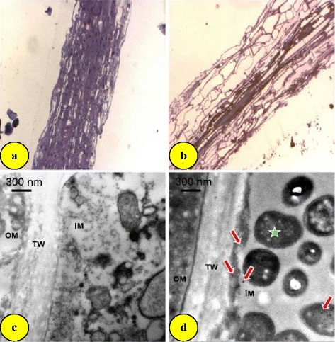
Fig. 3.

Images of barley (Hordeum vulgare) primary root tips. Light microscopic observation (magnification, ten times) of longitudinal sections of barley primary root tips of (a) control plants and (b) plants exposed to 10 μg mL−1 gold nanoparticles; TEM images of root cross-sections of (c) control plants and (d) plants exposed to 10 μg mL−1 gold nanoparticles. Bacteria (asterisk) and gold nanoparticles (arrows). OM outer matrix, IM inner matrix, TW thick wall [24]
