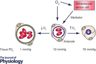
Figure 2. Schematic diagram of O2 signalling in the microcirculation .

Oxygen in the environment of arterioles can act directly on the arteriolar wall or on cells in the lumen to produce a vasomotor effect (dilatation in the case of reduced , or constriction in the case of elevated ). Alternatively, changes in may be sensed by extravascular cells (parenchymal cells, mast cells, nerves, etc.), a mediator produced, which then acts on the vessel wall to produce the appropriate vasomotor effect. The values shown below the cross section of the arterioles refer to tissue in a superfused, intravital preparation measured at the midpoint between the venous ends of two capillaries as reference values, only.
