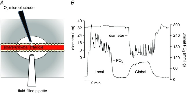
Figure 3. Local increases in have no effect on arteriolar tone .

Data shown are modified from Jackson (1987). A, schematic diagram of a segment (1–8 mm) of an arteriole in a hamster cheek pouch from which the parenchyma has been surgically removed (aparenchymal arteriole) to obviate effects of local changes on parenchymal and other extravascular cells. A Whalen‐type recessed tip O2 microelectrode was inserted through the wall of the vessel into the lumen as shown to monitor luminal . A temperature‐controlled micropipette filled with PSS equilibrated with varied (fluid‐filled micropipette) was positioned opposite the O2 microelectrode. Pressurization of the fluid‐filled micropipette ejected the O2‐equilibrated solution onto the surface of the arteriole to produce a local change in . The entire cheek pouch preparation was superfused with PSS, the of which could be varied to produce global changes in that affected both the aparenchymal arteriole and all other vessels and cell types in the cheek pouch. The diameter of the arteriole was measured by intravital video microscopy. B, results from a typical experiment in which either local increases in were produced using the fluid‐filled pipette (Local) or global increases in were produced by changing the of the entire superfusate (Global) (replotted data are from Fig. 3 in Jackson, 1987). Local increases in that effectively changed the across the wall of the arteriole had no significant effect on arteriolar diameter, whereas a global increase in produced sustained vasoconstriction. These data suggest that components of the arteriolar wall (endothelial cells, smooth muscle cells or perivascular nerves) are not the sensor cells responsible for arteriolar O2 reactivity in the hamster cheek pouch. See Jackson (1987) for details.
