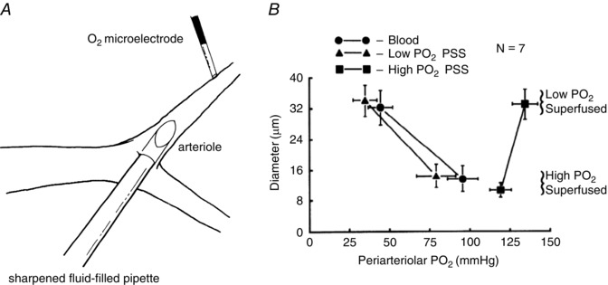
Figure 5. Perfusion of arterioles in situ with solutions equilibrated with high has no effect on arteriolar tone .

A and B are reproduced from Jackson (1987). A, schematic diagram of a hamster cheek pouch arteriole in which a sharpened fluid‐filled pipette has been inserted through the wall of an arteriole allowing perfusion of the arteriole with PSS equilibrated with varied . The entire cheek pouch preparation was superfused with PSS to allow global changes in . B, summary data (means ± SEM). Perfusion of the arterioles with solutions equilibrated with high or low had no significant effect on arteriolar diameter. Only when the global was elevated via the superfusate did the arterioles constrict (compare low superfusate points with high superfusate points). These data suggest that components of the arteriolar wall (endothelial cells, smooth muscle cells or perivascular nerves) are not the sensor cells responsible for arteriolar O2 reactivity in the hamster cheek pouch. See Jackson (1987) for details.
