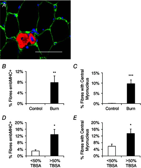
Figure 2. Skeletal muscle regeneration is elevated following burn injury .

A, representative immunohistochemical image demonstrating embryonic myosin heavy chain (embMHC) positive muscle fibres (red), laminin (green) and DAPI (blue). Scale bar = 50 μm. B, quantification of the frequency of embMHC+ fibres, expressed as mean percentage of fibres positive for embMHC ± SEM. C, quantification of the frequency of centrally nucleated muscle fibres, expressed as mean percentage of fibres displaying a central myonucleus ± SEM. D, comparison of the frequency of embMHC+ fibres in burn patients with <50% or >50% total body surface area (TBSA) burn percentage, expressed as mean percentage of fibres positive for embMHC ± SEM. E, comparison of the frequency of centrally nucleated muscle fibres in burn patients with <50% or >50% total body surface area (TBSA) burn percentage, expressed as mean percentage of fibres displaying a central myonucleus. n = 12 (Control) and 12 (Burn) subjects. Significant effect of burn injury: * P < 0.05, ** P < 0.01, *** P < 0.001.
