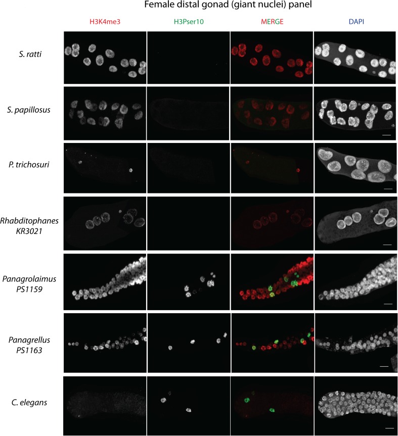
Fig. 6.
H3K4me3 and H3Pser10 staining patterns in females of different nematode species. H3K4me3 (in red) and H3Pser10 (in green) antibody stainings (with the individual channels separated according to color and labeled on top) in seven different (adult female) nematode species in the distal part of their gonads (distal tip is to the left for each). This region consists of giant nuclei in Strongyloides species. Note the similarity in gonad organization in S. ratti, S. papillosus, P. trichosuri, and Rhabditophanes KR3021, and between Panagrolaimus PS1159, Panagrellus PS1163, and C. elegans. For nematodes with a gonad organization similar to S. ratti, note the lack of H3Pser10 staining (in green) in the distal gonad, whereas its presence in species with a gonad organization similar to C. elegans. Scale bar 10 μm (note: adults are approximately 28–30 h post-culturing for Strongyloides species, but for other nematodes were young females carrying eggs in their uteri)

