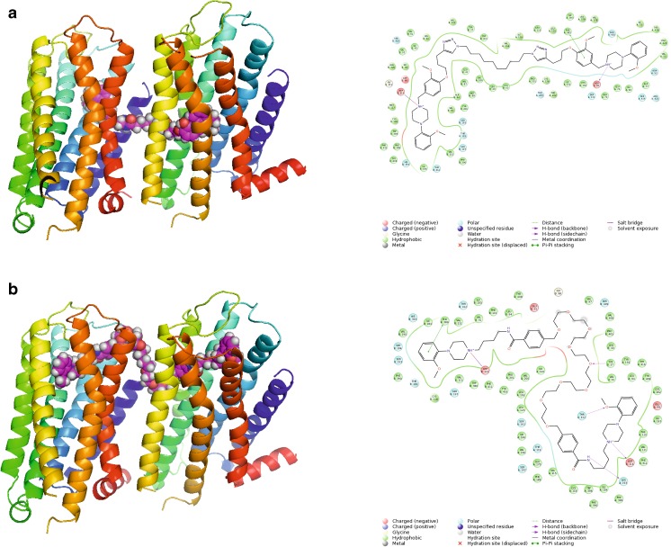
Fig. 9.
Different types of poses for bigger ligands. a Compound 4c crosses the membrane to direct the other monomer (improbable pose). b Compound 5j interacts with two orthosteric sites (a probable pose). Protein shown in ribbon representation, colored in spectrum mode. Ligands shown as spheres with magenta carbon atoms. 2D interaction maps also presented

