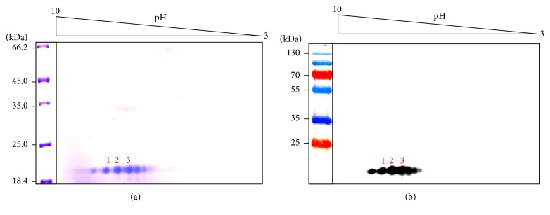
Figure 2.
Two-dimensional gel electrophoresis of the purified rLrIL-10. The purified rLrIL-10 was resolved by isoelectric focusing using carrier ampholytes (pH 3–10, shown on top) followed by SDS-PAGE (12%). Panel (a) shows coomassie brilliant blue stained gel. The spots 1–3 were subjected to MALDI-TOF-MS (shown in Figures 1–3 in Supplementary Material). (b) Western blot analysis of identical gel shown in Panel (a) using anti-rLrIL-10 antibodies. kDa indicates migration of the protein markers.

