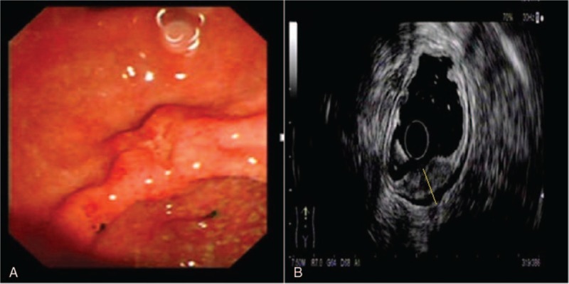
Figure 1.

Correct diagnosis of T staging in a patient with well differentiated gastric cancer. (A) Endoscopic image of the gastric cancer showing an ulcer located in the anterior wall of the antrum; (B) EUS image of the lesion showing a 13-mm thick hypoechoic lesion spreading from the mucosal to muscularis propria layers with an intact serosa layer (dotted line). Surgical resection confirmed well differentiated gastric cancer infiltrated to the muscularis propria layer.
