Figure 3.

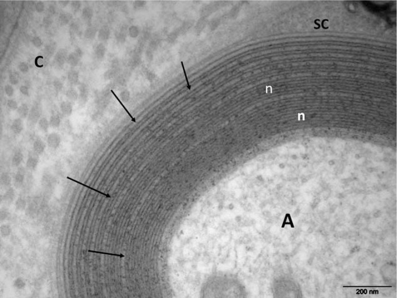
Electron micrograph, transverse section. At high magnification numerous regular widenings of the myelin lamellae are well seen (arrows). Otherwise, the myelin compaction is normal (n). A = axon, C = collagen, SC = Schwann cell cytoplasm.

Electron micrograph, transverse section. At high magnification numerous regular widenings of the myelin lamellae are well seen (arrows). Otherwise, the myelin compaction is normal (n). A = axon, C = collagen, SC = Schwann cell cytoplasm.