
Figure 3.
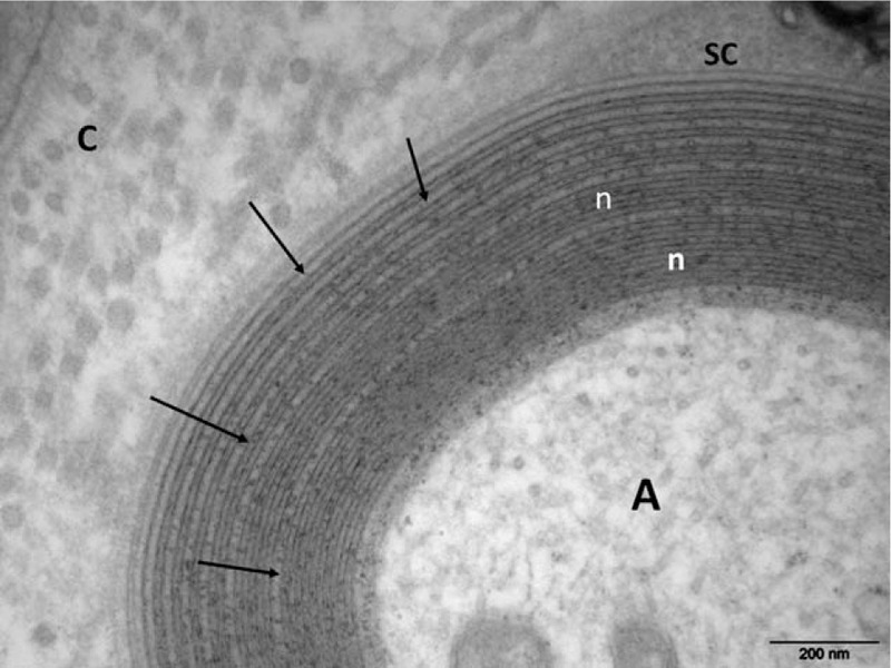
Electron micrograph, transverse section. At high magnification numerous regular widenings of the myelin lamellae are well seen (arrows). Otherwise, the myelin compaction is normal (n). A = axon, C = collagen, SC = Schwann cell cytoplasm.
Official websites use .gov
A
.gov website belongs to an official
government organization in the United States.
Secure .gov websites use HTTPS
A lock (
) or https:// means you've safely
connected to the .gov website. Share sensitive
information only on official, secure websites.
Electron micrograph, transverse section. At high magnification numerous regular widenings of the myelin lamellae are well seen (arrows). Otherwise, the myelin compaction is normal (n). A = axon, C = collagen, SC = Schwann cell cytoplasm.