
Figure 4.
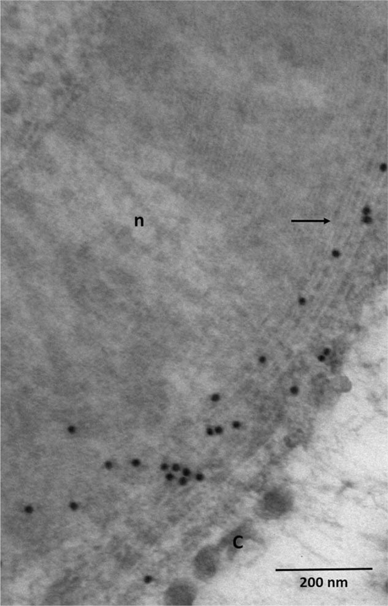
Immunoelectron micrograph, transverse section. Presence of IgG (black granules) at the level of widenings of the myelin lamellae (arrows). C = collagen, n = normally compacted myelin.
Official websites use .gov
A
.gov website belongs to an official
government organization in the United States.
Secure .gov websites use HTTPS
A lock (
) or https:// means you've safely
connected to the .gov website. Share sensitive
information only on official, secure websites.
Immunoelectron micrograph, transverse section. Presence of IgG (black granules) at the level of widenings of the myelin lamellae (arrows). C = collagen, n = normally compacted myelin.