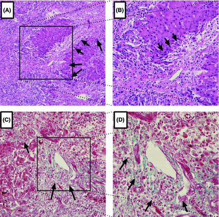
Figure 1.

Representative Light Photomicrographs of liver Tissue Showing Collagen Deposition. Original Magnification 100(A, C) or 200(B, D). Histopathological examination of the liver explants by H&E (A, B), and Masson(C, D) staining revealed collagen deposition, as highlighted by arrowhead.
