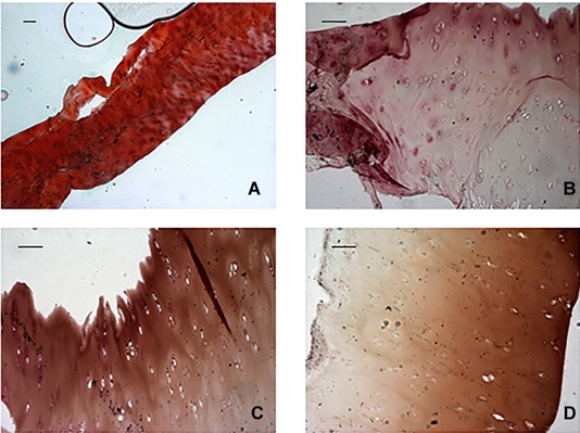
Figure 1.

Representative images of Safranin‐O staining. (A) Normal cartilage (horizontal sections of the central portion of menisci) displayed strong orange‐red staining, indicating a completely preserved cartilage. In contrast (B, C, D), AKU menisci (horizontal sections of the central portion of menisci) displayed moderate orange‐red staining, indicating advanced cartilage destruction. Scale bar = 100 μm.
