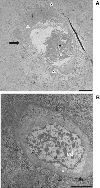
Figure 6.

TEM observation of AKU cartilage. Samples from Patient 1 (Table I) were from the superficial zone of cartilage (see Fig. 1B,a). Chondrocytes were phenotypically ascribed to those with phenotype, though being located in a non‐pigmented area. Extrusion of cellular material into the lacunae into extracellular space, typical feature in chondroptosis, was evident (A, arrow). The disintegration of cell in chondroptosis is a combination of digestion in autophagic vacuoles and of the release of resultant cell fragment into the extracellular space, as observable in part A (arrow heads). In this terminal stage of cell death, cytoplasm organelles were not recognizable and only some nuclear remnant is often visible (A and B). Chondroptotic remnants were disintegrated into vesicular detritus (B, vd). Bars A, B: 25 μm.
