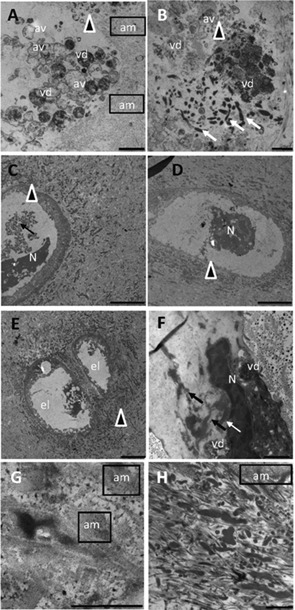
Figure 7.

TEM observation of AKU cartilage. Images offer an overview of AKU chondroptotic chondrocytes with secretory phenotype located in the deep zone and an ultrastructural analysis of extracellular matrix where collagen disaggregation (G, H) and diffuse pigment deposition (C–H) were evident. Cellular disintegration as the final stage of chondroptosis was evident in parts A, B, and E. The cell was totally fragmented into vesicles (vd: vesicular detritus) accumulated in the extracellular matrix, and the nucleus was barely visible (A, B). Cell remnants (arrowheads) were observable in parts C–F, where glycogen was absent, nucleus (N) was still visible but as a dense central region with some vacuoles and no trace of cytoplasm and organelles. In parts B and F condensed endoplasmic reticulum and Golgi were still visible (arrows), although very deteriorated, often forming compartments within which organelles were sequestered and digested (vd). This phenomenon of degradation also took place in autophagic vacuoles (A, B, av) and was followed by the expulsion of blebs into the lacunar space. In the nucleus, chromatin was patchy condensed and spread throughout the nucleus (E, F). Finally, the end stage of chondroptosis was an empty lacuna (C, el). Amyloid (am) was clearly visible in different images of AKU cartilage, as well as ochronotic pigment patchy dispersed in the degraded cartilage matrix (G, H). Samples were from Patient 2 (A, B), Patient 3 (C, D, H), and Patient 4 (E, F, G). Bars: A: 5 μm; B: 10 μm ; C, D, E: 25 μm; F: 10 μm; G: 2.5 μm; H: 5 μm.
