Figure 2.

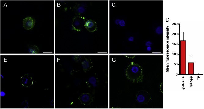
Uptake of rpdMspA and rpdApp into dendritic cells (DCs). Monocyte‐derived human DCs were incubated with FITC‐labelled rpdMspA (A), rpdApp (B) or TF (C). Cell nuclei were stained with Hoechst 33258. Co‐localization of labelled recombinant protein and nuclei (D) was determined using Zeiss LSM image examiner software. Mean fluorescence intensities were derived from ≥ 13 different fields from three independent experiments. Error bars: mean of values + SD. For inhibition, DCs were pre‐treated with mannan (1 mg ml−1; E), human transferrin (1 mg ml−1; F) or both inhibitors (G) for 30 min at 37°C before being incubated with FITC‐labelled rpdMspA. Cells were washed and fixed before analysis by confocal laser scanning microscopy. All images were scanned at a resolution of 1024 × 1024 pixels using the same laser and gains settings. The cells are representative of cells observed from three independent experiments. Scale bars = 10 μm.
