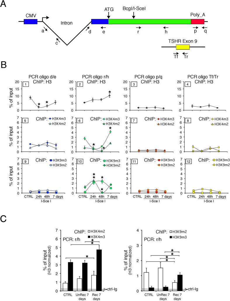
Figure 1. Spatial and temporal changes of histone H3 PTM after DSB at the GFP locus.
(A) Schematic diagram of DRGFP plasmid and a reference locus (TSHR exon 9). Structure of the DRGFP plasmid integrated as single copy in different locations in Hela cells. Primers a and c cannot be used in cells transiently transfected with I-SceI plasmid because they are also present in intron 1 of the I-SceI expression vector. (B) H3K4m2/3 and H3K9m2/3 content of GFP and the reference gene. DRGFP cells were transfected with I-SceI and characterized 24 h, 48 h and 7 days later. Cells were fixed and the chromatin analyzed by ChIP with the indicated antibodies. qPCR on each immunoprecipitate was carried out with the primers indicated in A. The specific antibodies are indicated at the top of each column. Each panel is identified by a numbered box in the upper left side.*P < 0.01 (t test) as compared with untreated control or basal. (C) H3K4m2/3 and H3K9m2/3 content in cells sorted 7 days after transfection. CTRL are cells transfected with a control plasmid; UnRec were GFP− cells sorted and separated from GFP+ Rec cells after I-SceI transfection. *P < 0.01 (t test) as compared with UnRec or CTRL. The detailed statistical analysis of the data shown in panels (B,C) is reported in Supplemental Statistical Tables 1 and 2.

