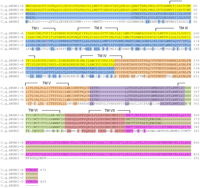
Figure 3. Alignment of the amino acid sequences of the four splice variants of C. gigas AKH receptor-1 (AKHR1).
The same color code is used as in Fig. 2, meaning that the amino acid residues highlighted with one color (Fig. 3) are coded for by the exon highlighted in the same color (Fig. 2). The transmembrane regions are indicated by TM I-TM VII. Also the AKHR2 is aligned with AKHR1-D. Identical amino acid residues between AKHR2 and AKHR1-D are highlighted in the colors corresponding to each exon of the AKHR1 gene. The highlighted F residue at position 46 of C. gigas AKH1-C was an S residue in the reference C. gigas genome sequence. Similarly, the highlighted T residue at position 110 of AKH1-C was an A residue in the reference C. gigas genome sequence. These changes are probably allelic variations. The cloned cDNA sequences were submitted to GenBank with the following accession numbers: C. gigas AKHR1-A, KM205066; C. gigas AKHR1-B, KM205067; C. gigas AKHR1-C, KM205068; C. gigas AKHR1-D, KM205069; C. gigas AKHR2, KM205070.

