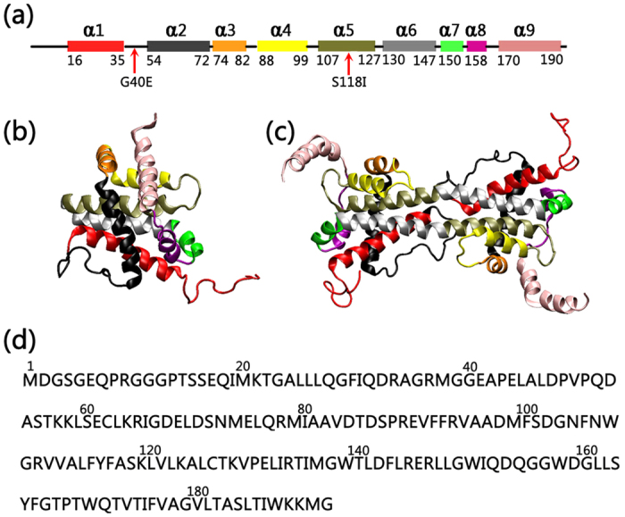
Figure 1.

(a) The locations of mutations are mapped on a schematic representation of the protein secondary structure. (b) The structures of the Bax monomer (lift) and dimer (right), and (c) the sequence of the Bax protein. α helices in Bax protein are represented by different colors. Color code for the helices: red (α1), black (α2), orange (α3), yellow (α4), tan (α5), silver (α6), green (α7), purple (α8) and pink (α9).
