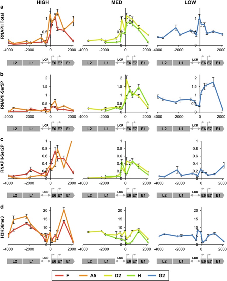
Figure 6.
Associations with RNAPII and H3K36me3. Levels of association of total RNAPII (derived from three biological replicates) (a), RNAPII-Ser5P (poised/paused) (three replicates) (b), RNAPII-Ser2P (active/elongating) (three replicates) (c) and H3K36me3 (two replicates) (d). In each graph, the y-axis shows the relative levels of enrichment, normalised to host control target regions (see Supplementary Table S1). The x-axis and underlying schematic show the region of the HPV16 genome analysed. In all panels, data are colour coded according to the key at the foot of the figure. Bars=mean±s.e.m.

