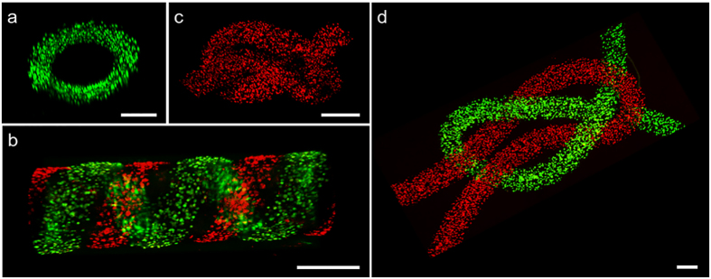
Figure 4. Fiber-based assemble of three dimensional structures.
Confocal images of (a) helical tube, (b) parallel helical tube, (c,d) different knots. Cells were labeled with Cell-tracker Green/Red. 32G needles were used in (a,b), and 30G (inner diameter 0.15 mM) needles were used in (c,d). Scale bar 200 μm.

