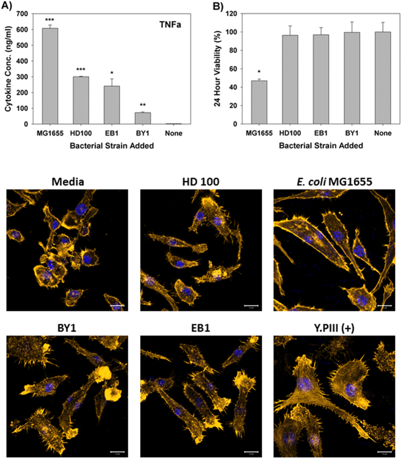
Figure 1. Response of RAW Macrophage 264.7 to predatory bacteria.
(A) ELISA was performed for TNF-α within Raw 264.7 mouse monocyte-macrophage cell cultures during a direct exposure to the predatory bacteria. The MOI was 1230 predators per single mammalian cell. E. coli MG1655 (MOI = 111) was used as a representative Gram-negative strain. The concentration of the inflammatory protein was measured 6 hours post-inoculation of the bacteria (n = 3). (*p < 0.05; **p < 0.01; ***p < 0.001). (B) MTT assay showing Raw 264.7 cell viability with the indicated bacterial treatments after 24 hours. The results are presented relative to the media control (100% viable). The mean values from three independent experiments are shown with the error bars representing the standard deviation. (*p < 0.05). (C) Impact of a 4-hour pre-treatment of Raw 264.7 mouse macrophage cells with predatory bacteria (1230 predators per single mammalian cell) on actin filament rearrangement. Raw 264.7 cells were cultured on collagen-treated 8-well chambered coverslips and fixed after 4 hours. The nuclei and actin filaments were stained with DAPI (blue) and Rhodamine-Phalloidin (gold), respectively. Composite images show the nuclei (blue) and actin (gold, false color added for better resolution). The formation of prominent stress fibers throughout the cytoplasm was only seen when the Raw 264.7 cells were exposed to Yersinia pseudotuberculosis YPIII (MOI = 111). Images are representative of n = 3 independent experiments. Scale bar = 10 μm.

