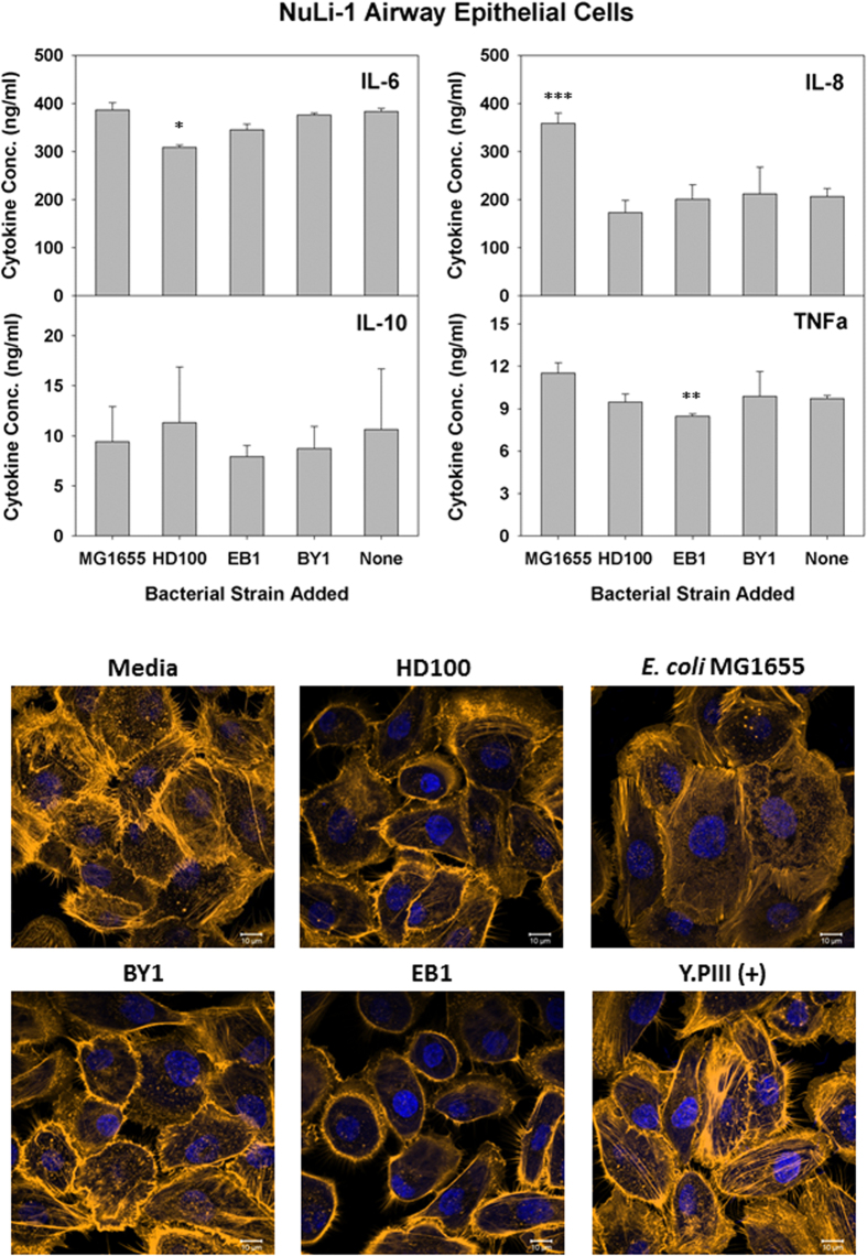
Figure 2. Induced inflammatory protein profile in response to predatory bacterial exposure to human alveolar epithelial NuLi-1.
(Upper Panels) ELISA assays were performed for human pro-inflammatory cytokines IL-8 and TNF-α (left panels), the dual functioning IL-6 and the anti-inflammatory IL-10 (right panels) in response to predatory bacteria (1230 predators per single mammalian cell) in NuLi-1 human lung epithelial cell cultures relative to the untreated media controls. E. coli strain MG1655 (111 bacteria per single mammalian cell) was used as a representative Gram-negative strain. Inflammatory proteins were measured 6 hours post-inoculation of the bacteria (n = 3). The mean values from three independent experiments are shown with the error bars representing the standard deviation. (*p < 0.05; **p < 0.01; ***p < 0.001). (Lower Images) Predatory bacteria induced no early cytoskeletal changes in the alveolar epithelial NuLi-1 cells. Impact of a 4-hour pre-treatment of NuLi-1 cells with predatory bacteria (1230 predators per single mammalian cell) on actin filament rearrangement. NuLi-1 cells were cultured on collagen treated 8-well chambered coverslip and fixed after 4 hours. Nuclei were stained with DAPI (blue) and actin filaments with Rhodamine-Phalloidin (red), and fluorescent confocal microscopy was performed. The composite images show nuclei (blue) and actin (gold, false color added for better resolution). Images are representative of n = 3 independent experiments. Scale bar = 10 μm.

