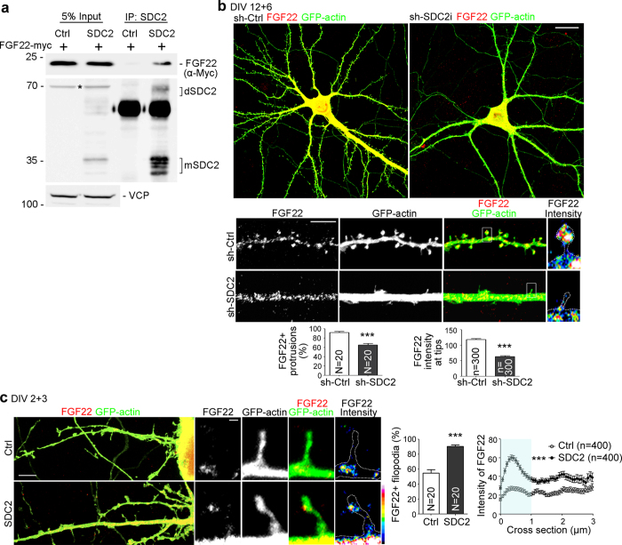
Figure 3. SDC2 facilitates FGF22 targeting to dendritic filopodia and spines.
(a) Coimmunoprecipitation of SDC2 and FGF22-myc. Neuro2A cells were cotransfected with indicated constructs and subjected to immunoprecipitation and immunoblotting using the indicated antibodies. Because of heparan sulfate modification and SDS-resistant dimerization via the transmembrane domain, multiple SDC2 bands are present in SDS-PAGE. dSDC2: SDC2 dimer; mSDC2: SDC2 monomer. Asterisk indicates non-specific signal. (b) Knockdown of SDC2 in mature neurons reduces the synaptic distribution of FGF22-mCherry. Whole cells and enlarged dendrite fragments are shown as indicated. The heat maps of individual protrusion show the intensities of FGF22-mCherry. (c) FGF22-mCherry is distributed to the tips of SDC2-induced filopodia. The results of the percentages of FGF22-positive filopodia and line scanning from the tips of filopodia to dendritic shafts are shown. The fluorescence intensities at tips indicated by the light blue area in the chart were used for statistical analysis. Data represent the mean plus SEM. N, number of analyzed neurons; n, number of analyzed protrusions. Samples were collected from two independent experiments. ***P < 0.001. Scale bar: (b) whole cell, 20 μm; dendrite, 5 μm; (c) lower magnification, 10 μm; high magnification, 1 μm.

