
Figure 3. N-terminal mutant forms of KATNB1 display reduced interaction with dynein and disrupted mitotic spindle.
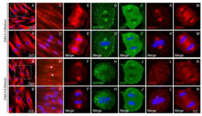
β-tubulin staining shows microtubule architecture to be intact in interphase dermal fibroblasts derived from patients and their parents (A–B). However, patient-derived cells display increased number of centrosomes (arrow) as seen by staining for γ-tubulin (C–D) and significantly disrupted and malformed mitotic spindle in anaphase (E–F). Patient fibroblasts also show reduced localization of KATNB1 (G–H), LIS1 (I–J) and dynein (K–N) to the mitotic spindle and spindle poles. Panels marked with a prime (′) show merged images of primary antibody and DAPI (blue) staining (A′–N′). All confocal images were captured using identical settings.