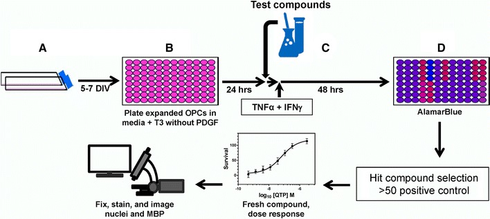
Fig. 4.

Flow scheme of the cytokine OL protection assay. A primary O4-positive OPCs were isolated for P6–P7 neonatal rat brains. A OPCs were expanded in flasks for 5–7 days in defined media supplemented with PDGF. B Expanded OPCs were plated in 96-well plates in defined media supplemented with T3, but without PDGF. C Test compounds were added following a 24-h incubation period followed 1 h later by addition of cytokine challenge (1 ng/ml TNFα+10 U/ml IFNγ). D After 48-h culture, alamarBlue® (AB) is added and AB fluorescence used to determine viability and metabolic activity of the cells. After selection of hit compounds (>50 % of the positive control, 1.1 µM QTP), fresh compound from a new source was obtained and the dose response relationship and EC50 values of each compound were determined. Following AB read out of the dose response, cells were fixed, immunostained, and images quantified to determine OL differentiation (MBP expression)
