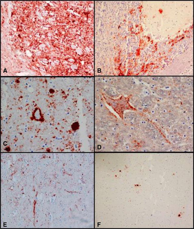
Figure 3.

IHC labelling of PrP CWD using F89/160.1.5 and 2G11 mAb. A Obex, intense coarse particulate labelling in the DMNV ×200. B Cerebellum, patchy labelling in the granular layer and stellate in the molecular layer ×200. C Ventral midbrain, scattered granules or accumulations of PrPCWD that appear as plaques ×400. D Ventral midbrain, neuronal and axonal labelling ×400. E Ventral midbrain, linear type labelling ×100. F Dorsal midbrain, sparse immunolabelling, plaque-like accumulation of PrPCWD ×100.
