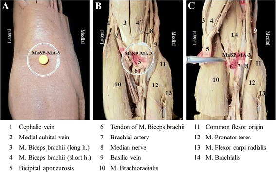
Fig. 3.

The visual example of the anatomical structures as marked by the presence of the acrylic color inject at the MaSP-MA-3, with its corresponding structures in the circular area. The anterior of the right elbow shows anatomical structures from superficial to deep layer. a The 1-inch circular area determines the anatomical structures of point. b Structures deep to point on superficial layer after removed skin and superficial fascia. c Structures in deep layer after flapping M. biceps brachii and aponeurosis
