
Figure 1. The Roles of Perivascular Macrophages in Tumor Progression.
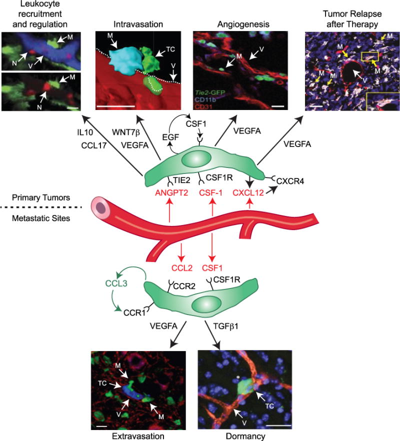
In primary tumors. Recruitment and regulation of other tumor-promoting leukocytes – the two images, with and without vessels (blue) included, show that neutrophils (red, N) extravasate in inflamed tissues in close proximity to perivascular (PV) macrophages (green, M). [Reprinted with permission: Abtin et al., 2014.] Intravasation of tumor cells: the images show a triad of a PV TIE2+VEGFA+ TAM (blue, M), cancer cells (green, TC), and endothelial cells (red). [Reprinted with permission: Harney et al., 2015.] Angiogenesis stimulation: the image shows TIE2+ TAMs (green, M) located near blood vessels (red, V) in tumors. [Reprinted with permission: De Palma et al., 2003.] Relapse of tumors after therapy: the images show a subcutaneous Lewis lung carcinoma after treatment with cyclophosphamide (TIE2+ blood vessels [red, V]; TIE2+MRC1+ TAMs [white/pink, M]; and cell nuclei [blue]). Inset: a single, TIE2+MRC1+ TAM (white/red). [Reprinted with permission: Hughes et al., 2015.] In metastatic sites. Extravasation of cancer cells: the image shows a cancer cell (blue, TC), PV macrophages (green, M), and blood vessels (red, V) in the lungs of mice. [Reprinted with permission: Qian et al., 2009.] Dormancy: the image shows a dormant cancer cell (green, TC; white asterisk) located close to a blood vessel (red, V) in the brain. Cell nuclei are shown in blue. [Reprinted with permission: Ghajar et al., 2013.] Many of the above functions involve the release of soluble factors by PV macrophages (green), and often activated by factors expressed by neighboring endothelial cells (red). Scale bars, 20 μm.