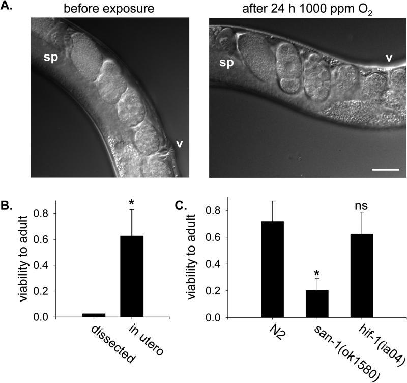
Figure 1. Embryos in utero suspend development and survive in hypoxia.
A. Nomarsky images of embryos in the uterus of a gravid adult C. elegans prior to and immediately after being exposed to 1000 ppm O2 for 24 h. Both pictures are of the same animal, and no embryos were fertilized or laid during exposure to hypoxia. v, vulva; sp, spermatheca of anterior gonad arm. Bar is 50 μm. B. Survival of embryos to adulthood after exposure to 1000 ppm O2 for 24 h. Dissected 2-cell embryos (n=40) were removed from the adult and exposed directly to hypoxia. Embryos in utero were laid by gravid adults exposed to hypoxia after return to normoxia (n=10 adults, 145 embryos). *, p<0.05. C. Viability of embryos exposed to 1000 ppm O2 for 24 h in utero. Gravid adult animals of each genotype were allowed to lay eggs exposed to hypoxia in utero upon return room air. The average number of embryos laid was 15.8±4.8 for N2 (n=18 adults), 9.4±3.6 for san-1(ok1580) (n=16 adults) and 10.7±3.8 for hif-1(ia04) (n=20 adults). The fraction of the embryos that hatched and developed successfully to L4/young adult was scored. Data are from two independent experiments. Error bars are standard deviation of the mean. *, p<0.05 compared to N2 controls; ns, not significant.

