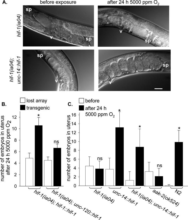
Figure 3. Neuronal HIF-1 expression rescues precocious engagement of diapause in hif-1(ia04) mutant animals.
A. Nomarsky images of embryos in the uterus of hif-1(ia04) (top panels) and hif-1(ia04); unc-14::hif-1 (HIF-1 expressed only in neurons; bottom panels) animals before and after exposure to 5000 ppm O2 for 24 h. The HIF-1 transgene contains a proline to alanine mutation that results in the constitutive stabilization of the protein, but does not otherwise affect its function [23]. v, vulva; sp, spermatheca. Bar is 50 μM. B. Embryo production over 24 h in 5000 ppm O2 was measured for groups of synchronized hif-1(ia04) mutant animals that carried extrachromosomal arrays with the indicated transgene. Animals that retained the array were identified after exposure to hypoxia (black bar) and compared to non-transgenic siblings that had had lost the array (white bars). The transgene constructs express HIF-1 from its native promoter (hif-1::hif-1) or in muscles (unc-120::hif-1). C. The number of embryos in the uterus was counted before (white bar) and after (black bar) 24 h exposure to 5000 ppm O2 for each genotype. Data from before and after exposure were compared: ns, not significant; *, p<0.05.

