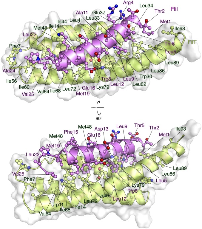
Fig. 5.
Solution structure of the FliTΔα4−FliIEN. FliTΔα4 is colored green, and FliIEN is colored pink. The solvent-exposed surface of FliT is represented in semitransparent light gray. Residues are shown in ball-and-stick configuration. The dashed lines denote hydrogen bonds. Two views are shown related by a 90° rotation about the x axis.

