Fig. S1.
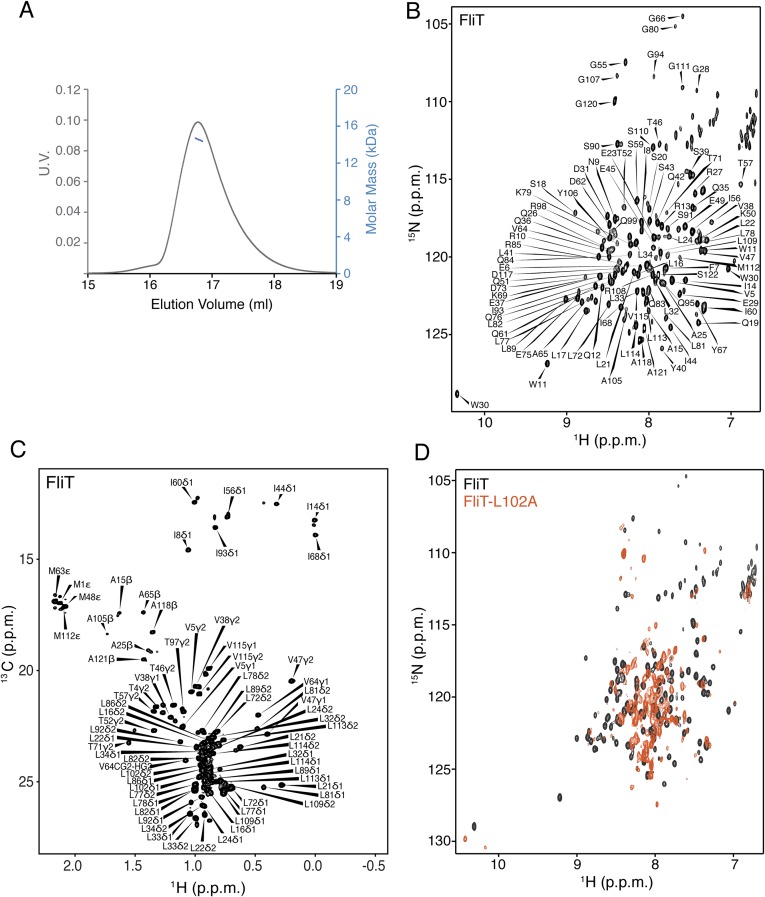
NMR characterization of FliT. (A) MALS of FliT shows that the protein exists as a monomer in solution. (B and C) 1H-15N heteronuclear single-quantum correlation (HSQC) (B) and 1H-13C HMQC (C) spectra of U-2H–,15N Ala-13CH3–, Met-13CH3–, Ile-δ1-13CH3–, Leu,Val-13CH3/13CH3–, and Thr-13CH3–labeled FliT. Resonance assignment is shown for the NH and methyl resonances. (D) 1H-15N HSQC spectrum of FliT-L102A (orange) superimposed on the spectrum of wild-type FliT (black).

