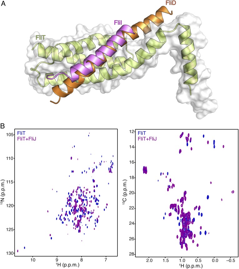
Fig. S9.
(A) Superposition of the FliT−FliIEN and FliT−FliDC structures demonstrates that FliI and FliD bind to the same surface on FliT. (B) 1H-15N HSQC (Left) and 1H-13C HMQC (Right) spectra of U-2H,15N Ala-13CH3, Met-13CH3, Ile-δ1-13CH3, Leu,Val-13CH3/13CH3, Thr-13CH3 of free FliT (blue) and in complex with FliJ (purple). NMR analysis shows that FliJ, FliD, and FliI bind to the same surface on FliT.

