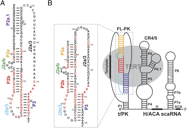
Fig. 1.
Medaka and human TR. (A) The secondary structure of the hFL-TR pseudoknot. The red letters indicate nucleotides with >80% sequence conservation among vertebrates. (B) Predicted secondary structure of mdTR containing the t/PK, CR4/5, and H/ACA domains. The FL-PK sequence and base pairs predicted by phylogenetic comparative analysis are shown in the box on the left. The 100%-conserved nucleotides identified in the five teleost fish TR are highlighted in red. TERT (gray ellipse) interacts with the t/PK and CR4/5 domains. The stems and loops are labeled and colored individually: P2a.1, purple; P2a, orange; P2b, red; P3, blue; J2a/b, green; J2a/3, black; and J2b/3, cyan.

