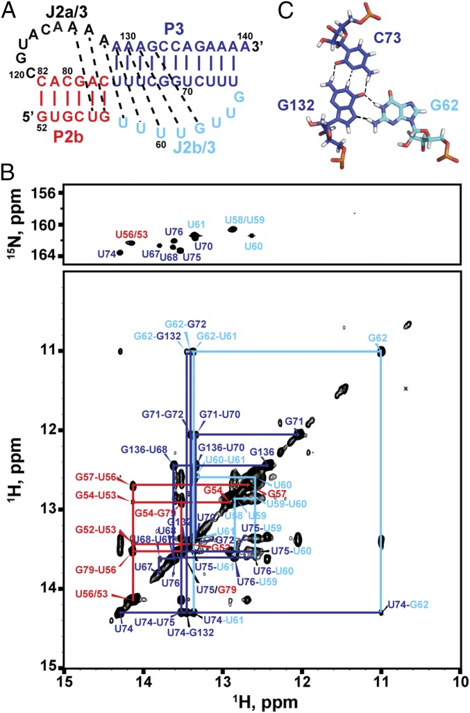
Fig. 2.
MdTR minimal (P2b–P3) pseudoknot. (A) Sequence and secondary structure of the minimal pseudoknot construct mdPK used in the solution NMR study. (B) Imino region of HNN-COSY (Upper) aligned with imino NOESY spectrum of mdPK (Lower). NOE cross-peaks of base pairs are connected and colored as in Fig. 1. (C) Stick representation of the G62–C73–G132 base triple. Secondary structure elements are colored as in Fig. 1.

