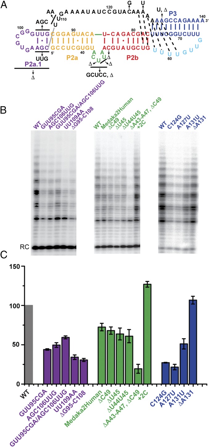
Fig. 4.
Telomerase activity assays of mdTR mutants. (A) Secondary structure of FL-PK with nucleotide substitutions indicated by arrows. (B) Effect of mdP2a1a, mdP2ab, and mdPK mutations (from left to right) on telomerase activity. Medaka TERT synthesized in RRL was assembled with full-length WT (first lane for each subgroup) or mutant mdTR. RC, recovery control. (C) Plot of the activity of each mutant relative to that of WT mdTR. The error bars indicate the difference or SD calculated from two or three independent assay reactions, respectively. Secondary structure elements and corresponding labels and bars in the plot are colored as in Fig. 1.

