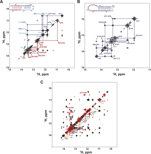
Fig. S6.
Comparison of the imino regions of H2O NOESY spectra of mdP2b, mdP3, and mdFL-PK. (A) mdP2b. (B) mdP3. (C) Superimposition of mdFL-PK (black) and mdP2b (red) spectra. The assignments of mdFL-PK are labeled along the diagonal using the color scheme in Fig. 1. Resonances from mdP2b are labeled in red to demonstrate the presence of a small amount of the P2b hairpin in mdFL-PK.

