Fig. 4.
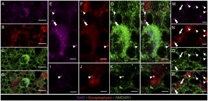
Confocal microscopy images of brain cryosections labeled with mouse anti-GAD (magenta), guinea pig anti-synaptophysin (red), and rabbit anti-NR1 (green), of the granular cell layer of cerebellum. (A–D) A GAD+ GoC (magenta, indicated in A) is represented, showing membrane staining for NR1 (green, C). (E–H) Magnifications of the GAD+ cell represented in A showing single (red, green, or magenta pixels for synaptophysin, NR1, and GAD staining, respectively) or multiple labelings marked with different labels: arrowhead for triple colocalization of GAD, synaptophysin, and NR1 signals; double arrowhead for colocalization of GAD and synaptophysin signals; and arrow for colocalization of synaptophysin and NR1 signals. (I–P) Taken from granular layer neuropil, different spots with high degree of labeling colocalization are indicated using the same label code used in E–H. [Scale bars: 50 μm (A–D) and 15 μm (E–P).]

