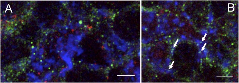
Fig. S6.
Confocal microscopy image of brain cryosections labeled with mouse anti-gephyrin (red), guinea pig anti-synaptophysin (blue), and rabbit anti-NR1 (green), of the granular layer of cerebellum. (A) The image reports a cerebellar glomerulus (synaptophysin+, bright blue staining) on the Right side and glomerular cell bodies (green background staining) on the Left side. In all these structures, NR1 immunoreactivity (bright green spots) and gephyrin immunoreactivity (bright red spots) never colocalize. (B) Arrows indicate synaptophysin/NR1++ spots in a glomerulus. No gephyrin+ structure is located close to synaptophysin/NR1++ spots. A semiquantitative evaluation on 10 randomly chosen glomerular structures could not find any instance of colocalization (0/82) between synaptophysin/NR1++ spots (i.e., presynaptic NR1+ structures) and gephyrin+ spots. (Scale bars: 15 μm.)

