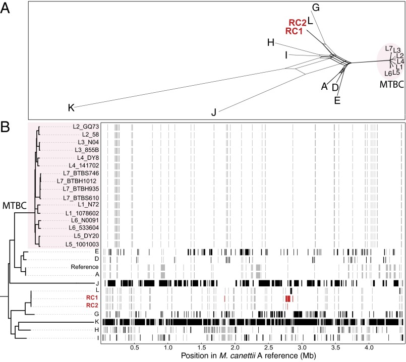
Fig. 2.
Recombinogenic vs. clonal strain cluster organization, distinguishing M. canettii and MTBC strains of lineages 1–7, respectively. Assessment of recombination among tubercle bacilli. (A) NeighborNet analysis of 9 M. canettii genomes and 16 MTBC genomes (representing each of the seven lineages), showing extensive recombination among the early branching tubercle bacilli relative to the MTBC. Relationships were inferred based on alignments of 53,603 variable nucleotides identified by whole genome comparisons against the M. canettii A reference sequence. (B) ClonalFrameML analysis of recombination of the same 27 genomes used in A above, showing the extent of recombination among M. canettii and the early-branching tubercle bacilli. The maximum likelihood phylogeny was inferred by using FastTree. Black horizontal bars indicate recombination events detected by the analysis in extant taxa. Red horizontal bars indicate recombination events detected in the laboratory-generated strains RC1 and RC2. Gray-shaded horizontal bars are inferred recombination events in a hypothetical common ancestor.

