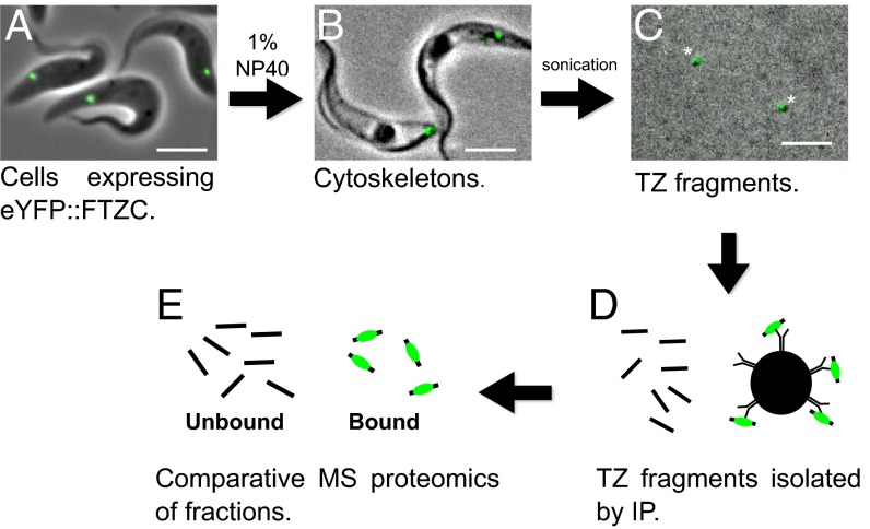
Fig. 1.
Isolation of transition zones. (A) Live cells expressing eYFP::FTZC at the TZ (green) were (B) treated with 1% Nonidet P-40 to extract the soluble material. (C) Washed cytoskeletons were sonicated to shear the TZs from other flagellar material. Tagged FTZC remained bound to the TZ at each step of the procedure (asterisk). (D) TZ fragments were isolated by anti-eYFP IP using Dynabeads and then (E) analyzed by mass spectrometry.

