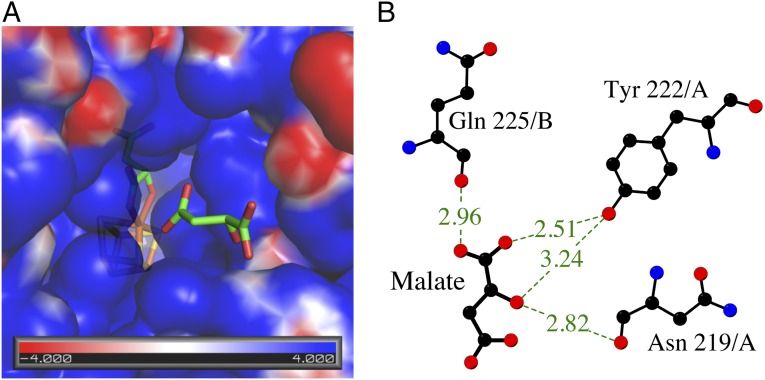
Fig. 3.
Substrate access to the active site of LmFH-2. (A) Electrostatic surface potential of the substrate-binding pocket showing the access of another S-malate (green) to the active site. The [4Fe-4S] cluster is shown in yellow (S) and orange (Fe). (B) Interactions between the second molecule of S-malate found in the positive cavity and N-terminal domain residues. The hydrogen bonds are shown as dashed lines. Image created with LigPlot (30).

