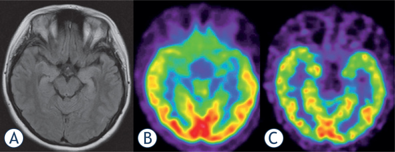
Figure 2.

18F-FDG PET (B)scan vs. 18F-Flumazenil PET (C)in a 19 year old female patient with bilateral hippocampal sclerosis as shown by MRI (A). Both PET modalities present low temporomesial uptake being larger on the left side, but benzodiazepinereceptor imaging appears sharper and presents a focal defect.
