Figure 5.
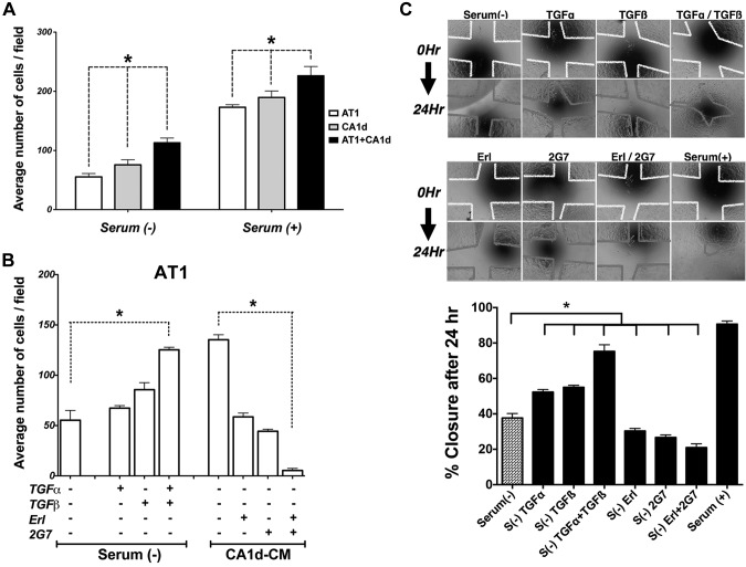
Effect of TGF-α and TGF-β on cell proliferation and motility. A) Cells were cultured under different conditions and their proliferation quantitated. Increased cell number was observed in presence of serum. Proliferation of mixed A/C cells significantly increased compared to AT1 or CA1d cells alone. *P < 0.05, 1-way ANOVA with Dunnett’s multiple comparison test. B) Addition of TGF-β alone or TGF-α + TGF-β increased proliferation of AT1 cells. When AT1 cells were exposed to CM from CA1d cells, the pan-TGF-β and EGFR inhibitors (2GT7 and Erl, respectively), alone or in combination, were able to significantly reduced growth of AT1 cells. *P < 0.05, 1-way ANOVA and multiple comparisons. C) Wound-healing assay was undertaken to assess motility of AT1 mixed with CA1d cells under different conditions. Photomicrograph of cells at beginning of experiment (top) and 24 h later (bottom). Colored lines indicate edge of scratch at beginning (0 h, yellow) and at end of experiment (24 h, red). Percentage of wound closure was calculated. Both inhibitors Erl and 2G7 had negative effect alone or in combination. Presence of serum had highest effect and almost completely closed gap between cells. *P < 0.05, 1-way ANOVA with Dunnett’s multiple comparison test.

