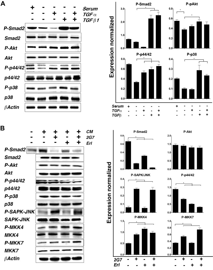
Figure 6.
Downstream effects of TGF-α and TGF-β activation in AT1 cells. A) Response of AT1 cells to TGF-α and/or TGF-β stimulation was evaluated. Increased activation of TGF-β pathway was observed in presence of serum, TGF-β, and combination of ligands compared to basal (serum −). Phosphorylation of p44/42 increased in presence of ligands compared to basal levels. Interestingly P-p38 MAPK had dramatic positive response only in presence of TGF-β. Right: average relative density of each band from 3 different experiments was quantitated and normalized against their corresponding control (total protein). Left: representative blot. *P < 0.05, 1-way ANOVA with Dunnett’s multiple comparison test. B) To address influence of secreted factors, AT1 cells were cultured in presence of CA1d CM and treated with TGF-β and/or TGF-α inhibitors 2G7 and Erl. TGF-β pathway was impaired with both inhibitors. While Akt pathway remained unchanged, P-p44/42 and P-p38 decreased with addition of inhibitors. Blocking TGF-β pathway significantly increased phosphorylation of SAPK/JNK and its MAPK2 activator MKK7, while MKK4 phosphorylation was increased in presence of both 2G7 and Erl. Right: quantitation of bands followed similar approach used in (A). Left: representative blot.

