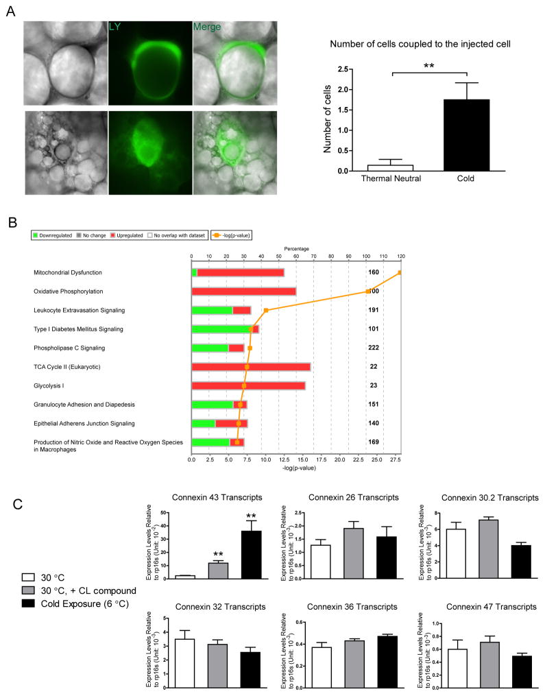
Figure 1. Increased Gap Junction Activity and Cx43 Expression during WAT Beiging.
A. Lucifer yellow coupling experiment, upper panel: adipocytes from mice kept at thermoneutrality; lower panel: beige adipocytes from mice kept at 6°C for 3 days. Quantification of the average number of cells coupled to the injected cell is shown on the right. n = 7 for adipocytes from mice housed at thermoneutrality, n = 10 for beige adipocytes. B. Pathway analysis of genes changed in the microarray study (cold treatment/thermoneutrality, mouse subcutaneous fat pad). C. Gene expression of various connexins in adipose tissue from mice at thermoneutrality, thermoneutrality and receiving CL316,243 treatment, or mice housed in the cold (n = 4–5). Results are shown as mean ± SEM, **p < 0.01.

