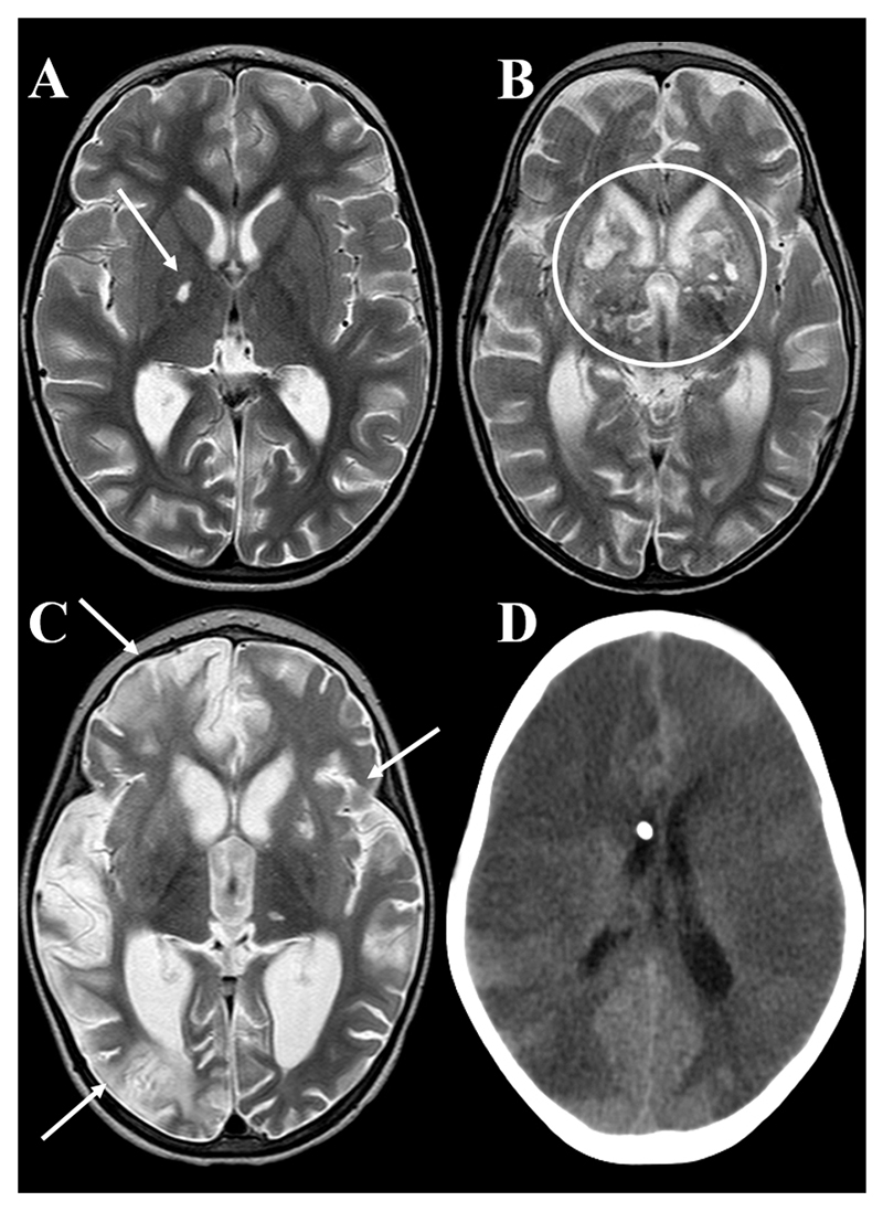
Figure 4. Infarcts.
Axial T2 weighted MRI showing A: small lacunar infarct involving the right globus pallidus (arrow), B: multiple bilateral MCA/ACA territory basal ganglia infarcts (circled), C: multiple hemispheric (arrows) and basal ganglia infarcts, D: axial uncontrasted CT showing global infarction in a patient who died before an MRI could be performed.

