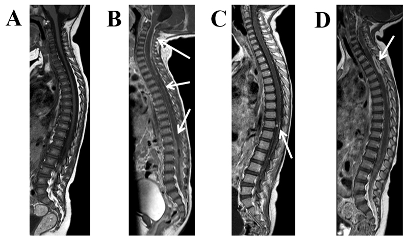
Figure 7. Pathology of the spine.
Sagittal T1 weighted MRI post-contrast in TBM: A: no enhancement, B: diffuse enhancement surrounding the cord and filling the thecal sac (arrows), C: cord and nerve root enhancement with an enhancing intramedullary tuberculoma (arrow), D: diffuse enhancement within the thecal sac and a focal intradural extramedullary plaque-like collection (arrow).

